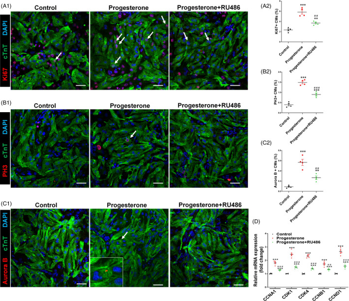
Figure 3.

Progesterone promotes CM proliferation in vitro in a progesterone receptor‐dependent manner. Cultured P7 CMs were treated with control vehicle (DMSO) or progesterone (10−7 M), alone or in combination with the progesterone receptor inhibitor RU486 (10−6 M). CM proliferation was analysed 24 h after progesterone stimulation. A‐C, CM proliferation was evaluated by Ki67, PH3 and Aurora B immunostaining. Representative images are shown in A1, B1 and C1, and quantification of percentages of Ki67+, PH3+ or Aurora B+ CMs is shown in A2, B2 and C2, respectively. (3000‐5000 cells were randomly selected and analysed for each group, and four independent experiments were conducted). Scale bars are 40 µm. D, The mRNA expression of cell cycle activators in P7 CMs was analysed by quantitative PCR (n = 5). (** P < .01, *** P < .001) vs Control; (## P < .01, ### P < .001) vs Progesterone. CM, cardiomyocyte
