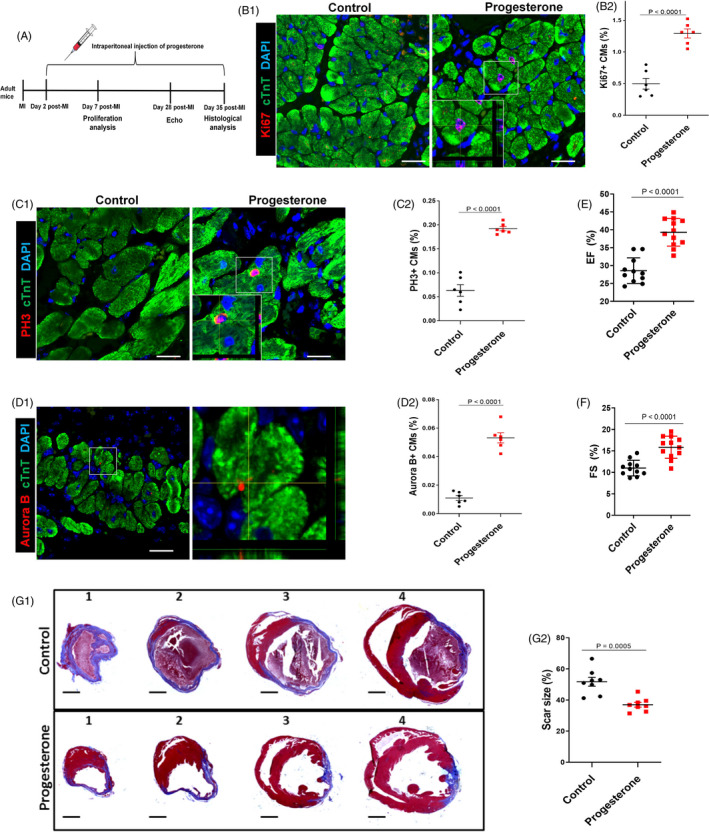
Figure 7.

Progesterone promotes adult CM proliferation and improves cardiac function after MI. A, Adult mice were subjected to MI by ligation of the left anterior descending coronary artery and intraperitoneally injected daily with progesterone (8 mg/kg) or control vehicle (corn oil) from day 2 to day 35 post‐MI. B‐D, Hearts were harvested at day 7 post‐MI; CM proliferation in the peri‐infarcted area was evaluated by Ki67, PH3 and Aurora B staining in CMs. Representative images with z‐stacking and quantification of percentages of those markers are shown (more than 5000 cells were randomly selected in each heart, and 6 mice were analysed in each group). Scale bars are 20 µm. E,F, Echocardiographic (Echo) analyses of the effect of progesterone on cardiac function were performed 28 days after MI. Quantitative analysis of left ventricular ejection fraction (EF) and left ventricular fraction shortening (FS) is shown (n = 11‐12). G, Hearts were harvested at day 35 post‐MI. Representative images (G1) and quantification (G2) of fibrotic scar of heart sections were analysed using Masson’s trichrome staining (n = 8). Scale bars are 1 mm. CM, cardiomyocyte; MI, myocardial infarction
