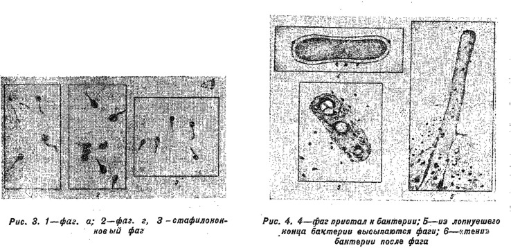
Figure 1.
Early drawings of bacteriophages destroying a bacterial cell, based on electron micrographs, to appear in Russian periodicals. Klein, op. cit. (note 35), p. 37, caption reads: ‘Drawing 3. 1—phage a; 2—phage g; 3—staphylococcus phage; Drawing 4. 4—phage attached to a bacterium; 5—phage pour out from the burst end of a bacterium; 6—“shadows” of a bacterium after phage’. (Reproduced courtesy of Nauka i Zhizn’.)

