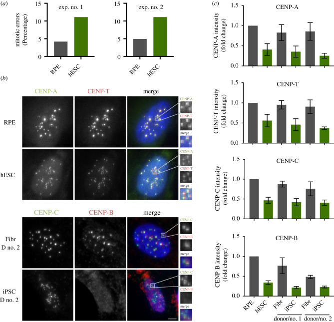
Figure 1.
Pluripotent stem cells have a weaker centromere than differentiated cells. (a) Quantification of mitotic errors in RPE and hESC, from two independent experiments. Cells were fixed and the frequency of mitotic errors in unperturbed cells was evaluated. (b) Differentiated (retinal pigment epithelium–RPE and fibroblasts from two independent donors–Fibr D no. 1 and Fibr D no. 2) and pluripotent stem cells (human embryonic stem cell line H9–hESC or iPSC lines reprogrammed from fibroblasts from donor no. 1 and donor no. 2–iPSC D no. 1 or iPSC D no. 2) were fixed and stained for CENP-A, CENP-T, CENP-C or CENP-B and counterstained with DAPI (blue). Representative immunofluorescence images from RPE and human embryonic stem cells (hESCs) are shown for CENP-A and CENP-T and representative images from fibroblasts and iPSC from donor no. 2 are shown for CENP-B and CENP-C. (c) Quantification of centromere intensities as shown in (a) for all cell types. Average centromere intensities were determined using automatic centromere recognition and quantification (CRaQ; see methods). The average and standard error of the mean of three replicate experiments are shown. Centromere intensities are normalized to those of RPE cells. Scale bar = 2 µm.

