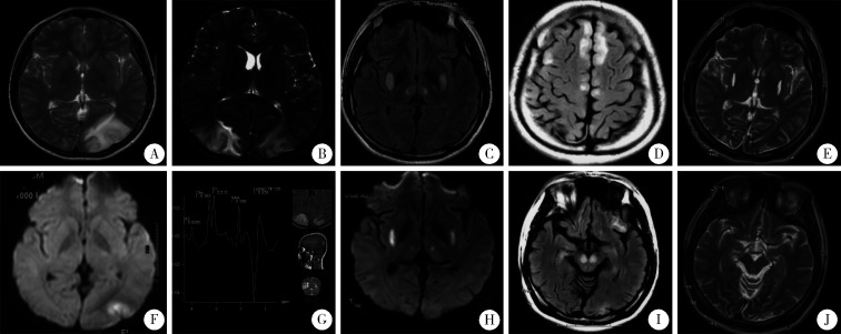
图1.
患者头颅MRI表现
Serial cranial MRI of the patient
At age 10 (A to D), left occipital cortex lesion with high signal on T2 and DWI imaging was shown (A, B); repeated MRI 20 days later the first MRI revealed a new lesion in the right occipital cortex with high-T2 signal (C), and magnetic resonance spectroscopy (MRS) showed lactate peak at the same time (D). At age 21(E to H), high-T2 and high-DWI signals were seen in bilateral putamen and thalami (E, F); bilateral new abnormal signals were in frontal cortex and cerebral peduncles when the condition worsened one month later (G, H). At age 24 (I, J), follow-up MRI showed regression of the bilateral putamen and thalami lesions, as well as complete disappearance of bilateral cerebral peduncles lesions (I, J).

