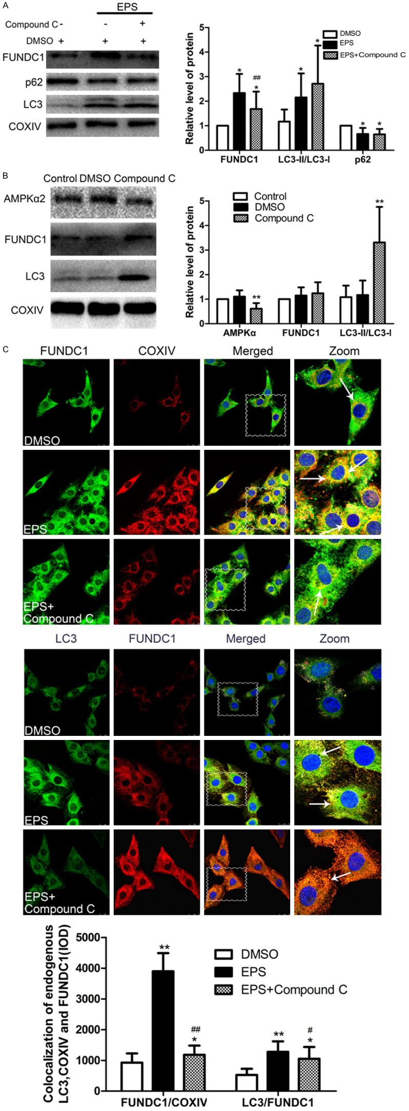
Figure 5.

FUNDC1 participates in EPS-induced mitophagy through the AMPK-ULK1 pathway. A. Western blotting of FUNDC1 and the indicators related to autophagy LC3 and p62. The values presented are means ± SD (n=6). *P<0.05, **P<0.01 in comparison to the control DMSO group; #P<0.05, ##P<0.01 in comparison with EPS alone. B. Western blotting of AMPKα2, FUNDC1 and LC3 in C2C12 cells with and without exposure to Compound C. The values presented are means ± SD (n=6). *P<0.05, **P<0.01, in comparison to the control DMSO group. C. Immunofluorescent staining of the FUNDC1-labeled outer mitochondria membrane (green or red) and co-localized with LC3 (green) and COXIV (red) (white arrows). Nuclei are stained blue with DAPI. (Scale bar: 25 µm). Quantitative IOD analysis of the co-localization of LC3, FUNDC1 and COXIV (means ± SD; (n=6). *P<0.05, **P<0.01 in comparison to the control DMSO group; #P<0.05, ##P<0.01 in comparison to treatment with EPS alone).
