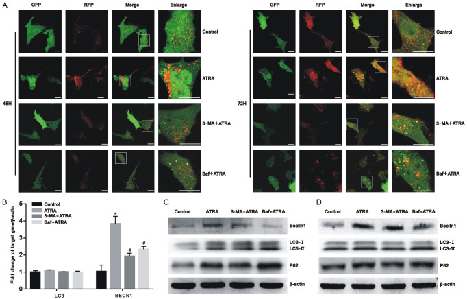
Figure 3.
Autophagy was successfully inhibited by 3-MA and Baf. Hepa1-6 and HepG2 cells were pretreated with 3-MA and Baf for 3 h before exposure to 10 μmol/L of ATRA. (A) After ptfLC3 transfection, the autophagic flux of Hepa1-6 cells was dynamically observed using a laser scanning confocal microscope after 48 h and 72 h of ATRA treatment. Scale bar = 20 μm; (B) After ATRA treatment, the mRNA expression of the BECN1 was upregulated and decreased in the presence of 3-MA and Baf, whereas LC3 showed no significant change among these groups in Hepa1-6 cells using real-time PCR. All results were obtained from three independent experiments. *P<0.05 vs. control group; #P<0.05 vs. ATRA group; (C, D) After ATRA treatment, the autophagy-related marker proteins LC3, Beclin1, and P62 were detected using western blot with β-actin normalization in Hepa1-6 cells (C) and HepG2 cells (D).

