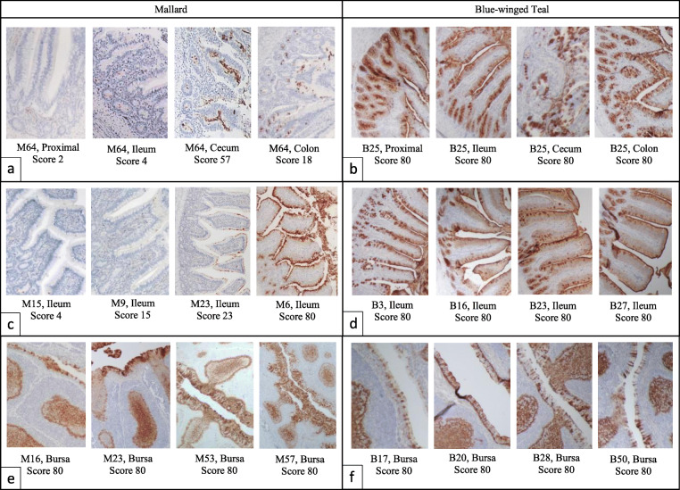
Fig. 4.
Lectin staining of mallard and blue-winged teal intestines and bursa of Fabricius. Lectin binding is positive where the brown colored stain is visible. The individual bird ID, tissue, and lectin score (villi enterocyte/epithelial cells) are given for each histological photograph. Scores were determined by averaging the scores for each field of view evaluated at 400x. Each field of view was given the following score: 0, no cells stained; 5, 1–10% of cells were stained; 35, 11–60% of cells stained; and 80, 61–100% of cells stained. Segments (a) and (b) show the range of lectin scores between sections of intestinal tissue in one individual (proximal represents duodenum or jejunum). Segments (c) and (d) show the range of lectin between individuals for the ileum tissue specifically. Segments (e) and (f) show lectin scores in the bursa of Fabricius. All photos were taken at 200x brightfield microscopy

