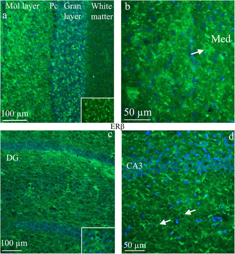
Fig. 3.

ERβ receptor distribution. a and b) ERβ receptor distribution in the cerebellum - the expression of ERβ was often observed in tiny loop-formations or short curvatures in areas close to the cell surface or intercellularly, reminiscent of extra cellular matrix distribution. In the cerebellum, the typical ERβ expression was found in the molecular (mol layer) and granular (gran layer) layers, but not in the Purkinje cells (Pc) and not in the white matter. Insert: staining of the granular layer. In Medial cerebellar nuclei (Med), occasional neuronal expression was observed (arrow). c and d) ERβ receptor distribution in the hippocampus - the dentate gyrus (DG) and CA3 are parts of hippocampus. Some ERβ positive pyramidal cells were found in DG (insert). In the other parts of hippocampus, staining resembling cell surface or extracellular matrix immunoreactivity was observed (arrows)
