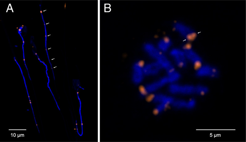
Fig. 4.

Chromosomal FISH of telomere repeats. Both panels show chromosomal fluorescent in situ hybridisation using probes against the canonical telomere sequence (TTAGGGx7). a In haploid spermatozoa, only one focus is visible for each of the six chromosomes (arrows), whereas two foci per chromosome (= 12) would be expected if telomeric repeats were present on both ends. b A metaphase figure shows chromatids joined at their centromeric ends, which lack probe signal, whereas probe is visible at the opposing ends of each sister chromatid (arrows)
