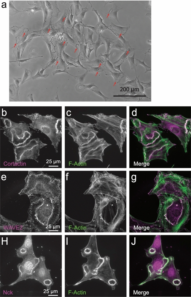
Figure 1.

The actin structures elicited with PDGF-BB treatment are bona fide CDRs. M28 mouse embryonic fibroblasts were serum-depleted in 0.2% FBS media for 12 h prior to addition of 20 ng/ml PDGF-BB for 6 m. (a) A low magnification phase contrase image shows numerous CDRs (select CDRs indicated with red arrows). (b–j) Immunofluorescence images demonstrating that F-Actin (labeled with phalloidin) (c,f,i) co-localizes with known CDR components cortactin (b), WASP2 (e) and Nck (h) as evidenced in the merge of these signals (overlap appears white) (d,g,j).
