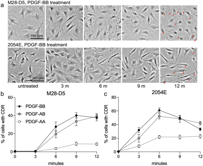
Figure 4.
Treatment with PDGF ligands stimulates CDR formation downstream of PDGFR activation. (a) Representative phase contrast images at various time points after PDGF-BB addition. Red arrows in the 12 m images highlight the presence of CDRs. (b,c) Quantification of the frequency of CDRs observed in M28-D5 fibroblasts (b) and 2054E melanoma cells (c) at various times after the addition of PDGF-AA (open circles), PDGF-AB (grey circles) or PDGF-BB (black circles).

