Figure 5.
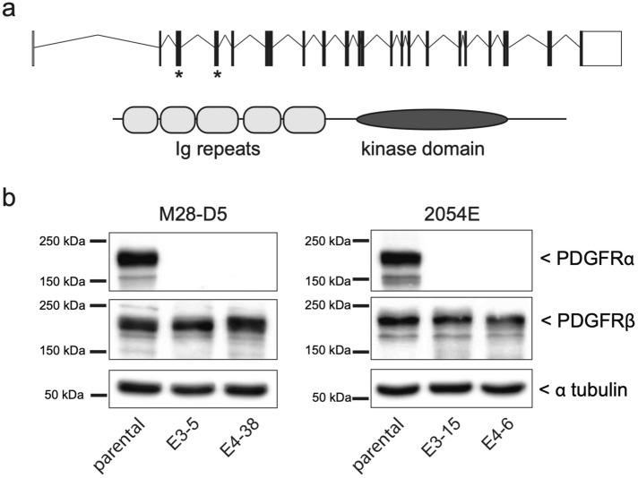
CRISPR/Cas9 gene editing disrupts PDGFRα expression. (a) Top: Schematic diagram of the mouse Pdgra gene, with exons depicted as boxes and introns as connecting lines. Asterisks indicate the exons targeted for CRISPR/Cas9 cleavage. Bottom: Domain structure of the mouse PDGFRα protein. (b) Cropped immunoblots showing the PDGFR expression profile of both the parental M28-D5 and 2054E cell lines, as well as the Pdgfra−/− cell lines derived from them. Cells were grown under standard culture conditions. Alpha-tubulin is used as a loading control. Full-length images of the cropped immunoblots are presented in Supplementary Figure S1.

