Figure 1.

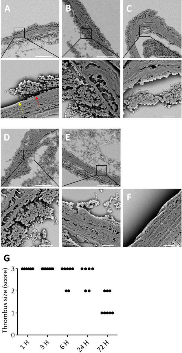
Temporary ligation of the carotid artery provokes consistent sub-occlusive thrombus formation in vivo. Scanning electron microscopy (SEM) images of thrombi (A) 1, (B) 3, (C) 6, (D) 24 and, (E) 72 h after temporary ligation of the carotid artery in control WT mice. (A–E) In all samples sub-occlusive platelet-rich thrombus formation is observed on the injured vessel wall. Damaged endothelium is indicated by a red arrow and undamaged endothelium by a yellow arrow. (F) No platelet adhesion was observed in absence of temporary ligation (3 mm upstream). Representative SEM images of 6 independent ligations are depicted. Scale bar is 80 μm, or 10 μm for zoomed images. (G) Thrombus size was scored by examining SEM images obtained from ligated carotid arteries that were collected at 1, 3, 6, 24 or 72 h after ligation. Scoring scale: 0 = no platelet adhesion, 1 = platelet monolayer, 2 = small thrombi (< 15 layers), and 3 = larger thrombi (> 15 layers).
