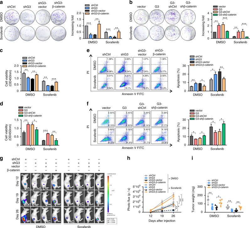
Fig. 5. Molecular deletion of Galectin-3-β-catenin signalling synergistically improves the antitumour effect of sorafenib in vitro and in vivo.
a–f After cell adhesion, DMSO or sorafenib (20 μmol/mL) were administered every two days for the colony formation assay, (a, b), CCK8 assay (c, d) and apoptosis assay (e, f) in Hep3B-vector, Hep3B-Galectin-3, Hep3B-Galectin-3-shControl, Hep3B-Galectin-3-shβ-catenin, Huh7-shControl, Huh7-shGalectin-3, Huh7-shGalectin-3-vector and Huh7-shGalectin-3-β-catenin cells. Scale bars in a and b, 5 mm. g, h Representative images and photo flux of tumour growth of Hep3B-vector, Hep3B-Galectin-3, Hep3B-Galectin-3-shControl, Hep3B-Galectin-3-shβ-catenin, Huh7-shControl, Huh7-shGalectin-3 Huh7-shGalectin-3-vector and Huh7-shGalectin-3-β-catenin cells in BALB/c nude mice treated with DMSO or sorafenib at day 12, 19, and 26. i Mice were sacrificed at day 27. The tumour weights were measured. Ctrl control, G3 Galectin-3. The results represent three independent experiments. *P < 0.05; **P < 0.01; ***P < 0.001.

