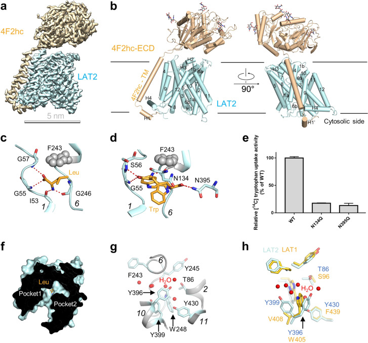
Fig. 1. Cryo-EM structure of the human LAT2–4F2hc complex.
a The overall Cryo-EM map of the human LAT2–4F2hc + Leu complex. LAT2 and 4F2hc are cyan and wheat, respectively. b Overall structure of the LAT2–4F2hc + Leu complex. The glycosylation moieties are shown as sticks. H helix, TM transmembrane domain, ECD extracellular domain. c The Leu-binding site. d The Trp-binding site. Trp substrate might have two binding positions. e Mutations of the Trp-binding residues lead to a decreased transport activity. Data are mean ± s.d. of three independent experiments. f The two separated pockets in LAT2. The pockets 1 and 2 are separated by the substrate. g The water distribution in the pocket 2 of LAT2. h The alignment of the second pocket of LAT1 with LAT2. The residues of LAT1 and LAT2 are shown as yellow and cyan, respectively.

