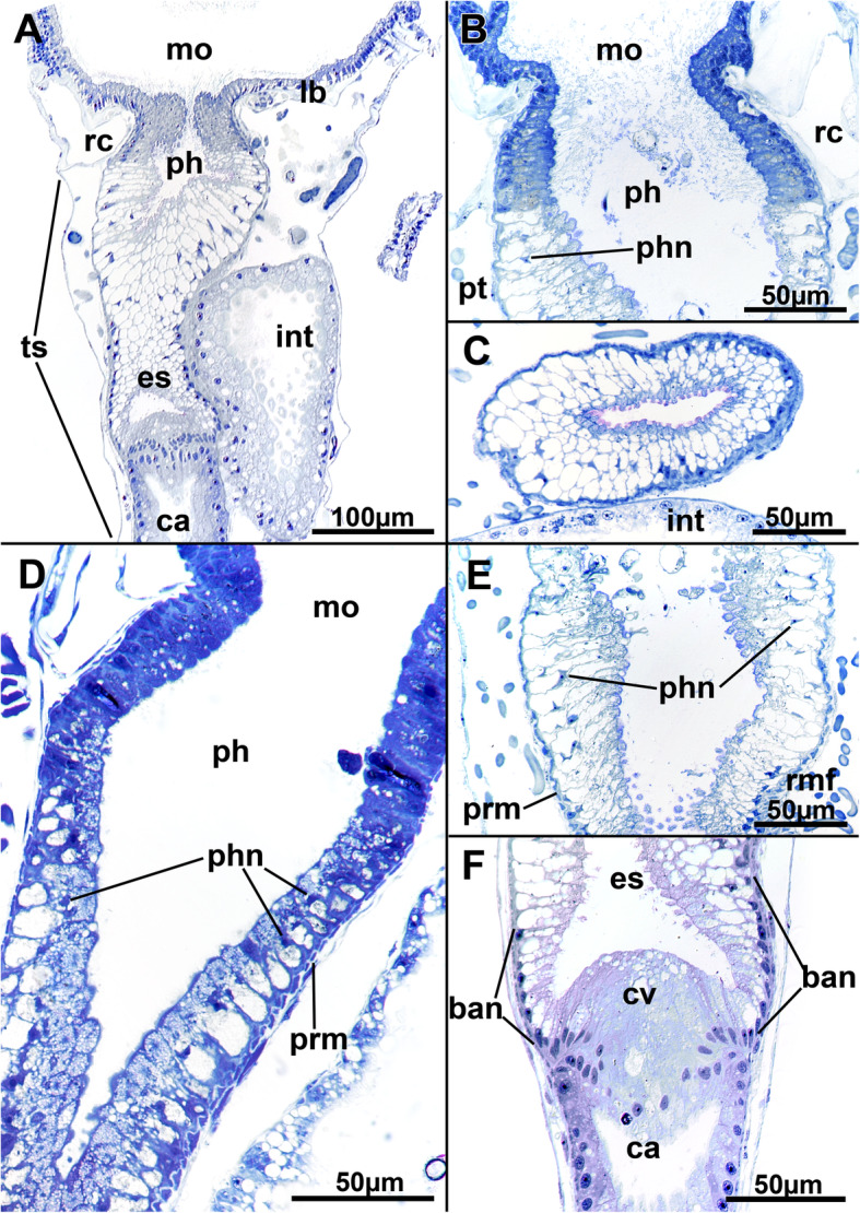
Fig. 10.

Histological details of the foregut of Stephanella hina. a Longitudinal section of an extended polypide showing the mouth opening, foregut until the cardia, and intestine. b Detail of a mouth opening and pharynx area in a zooid with retracted polypide. The nuclei of the pharynx appear pycnotic and are wedged between large vacuoles in the cells of the epithelium. c Cross-section of a pharynx showing the highly vacuolar appearance of its epithelium. d Longitudinal section of the mouth opening and pharyngeal area showing a high abundance of smaller vesicles in the apical part of the pharyngeal epithelial cells. e Detail of the lower part of the pharynx entering the esophagus. f The esophageal-cardia boundary is marked by the cardiac valve. On the esophagus, basally located nuclei are present in its cells. Abbreviations: ban - basal nuclei, ca - cardia, cv - cardiac valve, es - esophagus, int - intestine, lb. - lophophoral base, mo - mouth opening, ph - pharynx, phn - nucleus of the pharynx, prm - pharyngeal ring muscles, rc - ring canal, rmf - retractor muscle fibres, ts - tentacle sheath
