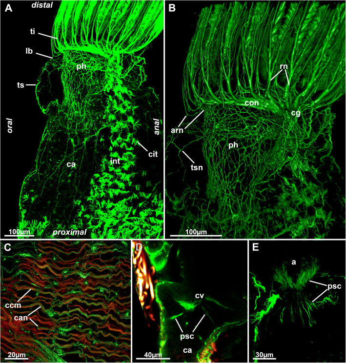
Fig. 16.
Nervous system of the digestive system of Stephanella hina. Confocal laser scanning microscopy stacks based on acetylated alpha-tubulin labelling. Volume renderings, projection or optical slices. Acetylated alpha-tubulin in green LUT, f-actin in glow LUT. a General overview of an extended polypide showing dense innervation of the foregut. Note also the dense ciliary bundles on the anal side of the intestine. b Details of the foregut shown in A. c Innervation of the caecum. d Optical section of the foregut-cardia transition showing few presumptive sensory cells projecting into the digestive epithelium. e Presumptive sensory cells in the lining of the intestinal wall close to the anus. Abbreviations: a – anus, arn – additional radial nerve, ca – cardia, can – caecum innervation, ccm – caecal muscles, cg – cerebral ganglion, cit – ciliary tufts, con – circum-oral nerve ring, cv – cardiac valve, int – intestine, lb. – lophophoral base, ph – pharynx, psc – presumed sensory cells, rn – radial nerve, ti – tentacle innervation, ts – tentacle sheath, tsn – tentacle sheath innervation

