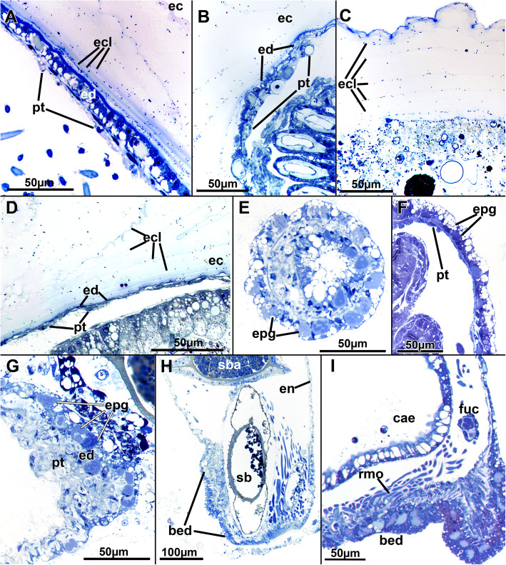
Fig. 2.
Histological details of the cystid wall of Stephanella hina. Semithin sections. a-d The ectocyst secretion consists of various concentric layers as seen by their granular stained borders. a Close to the endocyst, several distinct thin layers are discernible. The epidermis is prominent, while the peritoneum is not. b Body wall with thin epidermis and thicker, prominent peritoneum. c Distinct layers of the ectocyst are visible. The ectocyst stains only weakly, but its uppermost layer (top of image) is often more conspicuous. d Endocyst with extremely thin epidermis and peritoneum. e-g Epidermal glandular parts of the body wall. e Section of numerous translucent vesicles and other more distinctly stained vesicles. f Similar to E, longitudinal section. g Endocyst with a prominent peritoneal layer, probably containing lytic tissues and substance. h Longitudinal section of a zooid showing prominent glandular epidermis at the basal side (bottom of image) where the animal is attached to the substrate and the anchorage of the prominent retractor muscle fibres is located. i Detail of the basal epidermal attachment pad showing a high abundance of glandular vesicles. Abbreviations: bed - basal epidermis, cae - caecum, ec - ectocyst, ecl - ectocyst layers, en - endocyst, ed. - epidermis, epg - epidermal glands, fuc - funiculus, pt. - peritoneum, rmo - retractor muscle origin, sb - statoblast, sba - statoblast anlage

