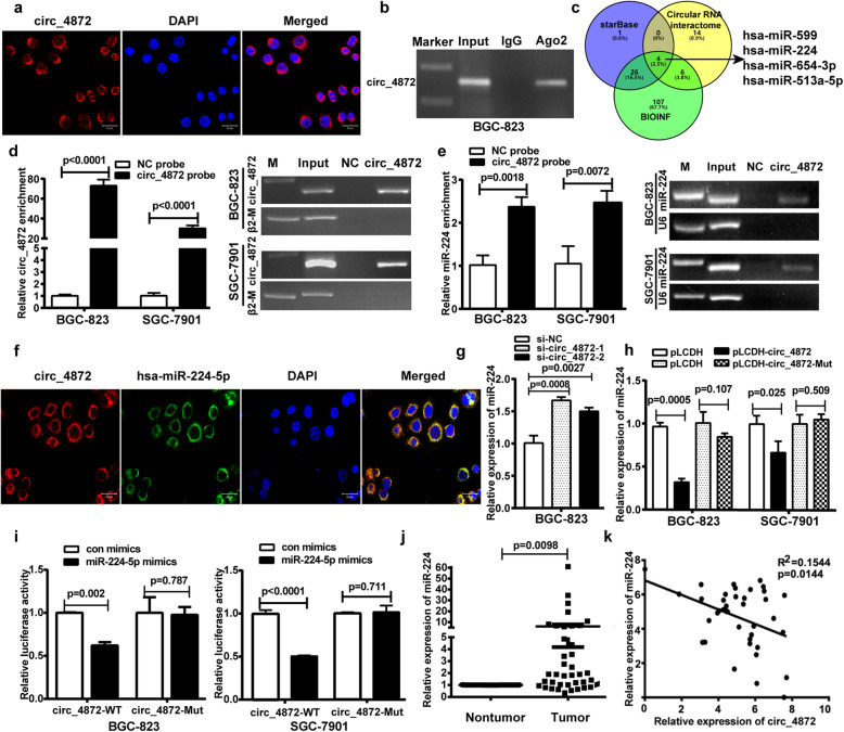
Fig. 3.
hsa_circ_0004872 acts as a “molecular sponge” for miR-224. a FISH experiment was used to detect the subcellular localization of hsa_circ_0004872 (circ_4872) in GC cells. b RIP analysis of hsa_circ_0004872 level in the immunoprecipitate of AGO2 antibody from GC cells. c Schematic diagram exhibiting the overlapping of different databases to predict the miRNAs with potential binding abilities of hsa_circ_0004872. d and e Lysates prepared from hsa_circ_0004872 overexpressing BGC-823 cells and SGC-7901 cells were incubated with biotinylated probes against hsa_circ_0004872 and then performed RNA pull-down assay. qRT-PCR and RT-PCR were used to determine the level of hsa_circ_0004872 (d) and miR-224 (e). f FISH analysis of the subcellular localization of hsa_circ_0004872 and miR-224 in GC cells. g qRT-PCR was used to determine the level of miR-224 in BGC-823 cells transfected with hsa_circ_0004872 siRNAs. h qRT-PCR was used to determine the level of miR-224 in BGC-823 and SGC-7901 cells transfected with hsa_circ_0004872 overexpression vector (pLCDH-circ_4872) or the mutated hsa_circ_0004872 overexpression vector (pLCDH-circ_4872-mut). i The wild-type (WT) or mutant (Mut) reporter constructs was cotransfected with control or miR-224 mimics into BGC-823 and SGC-7901 cells, and the dual luciferase activity was determined at 48 h after transfection. j qRT-PCR analysis of miR-224 expression in GC tissues and corresponding nontumor tissues with paired t-tests (n = 39, p = 0.0098). k Regression analysis of GC tissue showed a negative correlation between miR-224 and hsa_circ_0004872 (n = 39). All datas were the means ± SD

