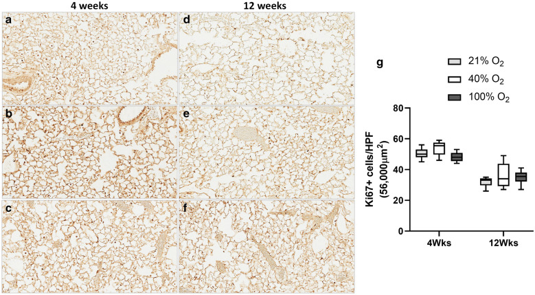
Fig. 2.
Ki67 Immunostaining. Staining for Ki67, a marker of cellular proliferation was performed on lung sections at four weeks (a 21%O2, b 40%O2 & c 100%O2) and 12 weeks mice (d 21%O2, e 40%O2 & f 100%O2) following neonatal oxygen exposure (Scale bar: 60 µm). Ki67 positive cells for nuclear staining was calculated at 400 × resolution (56,000 µm2 area) in all the groups (g five sections/mice; 6 mice/group; 21%O2—grey bars; 40%O2—white bars; 100%O2—black bars). The number of Ki67 positive cells demonstrated a significant correlation over time but not with the oxygen groups (p < 0.001, Two-way ANOVA). On multiple comparisons, there was no significant difference among the oxygen groups

