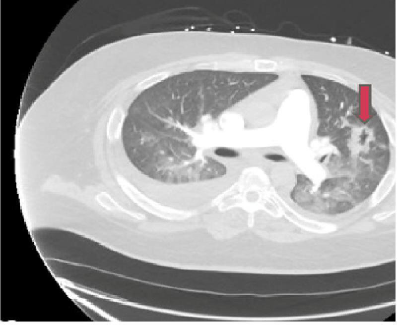
Figure 1.

CT angiography of the chest showing pulmonary vascular congestion and focal consolidation in the left upper lobe. The red arrow points to the cavitating lesion in the left lung field, which on biopsy was a non-specific inflammation and fibrosis.
