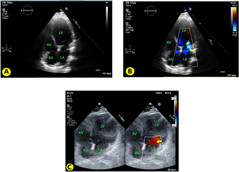
Figure 2.
(A) TTE images with apical four-chamber view without colour; (B) TTE with apical four-chamber view with colour Doppler showing mitral regurgitation (yellow arrow); (C) Follow-up TTE 3 months after discharge, showing decrease in mitral regurgitation (yellow arrow). LA, left atrium; LV, left ventricle; RA, right atrium; RV, right ventricle; TTE, transthoracic echocardiography.

