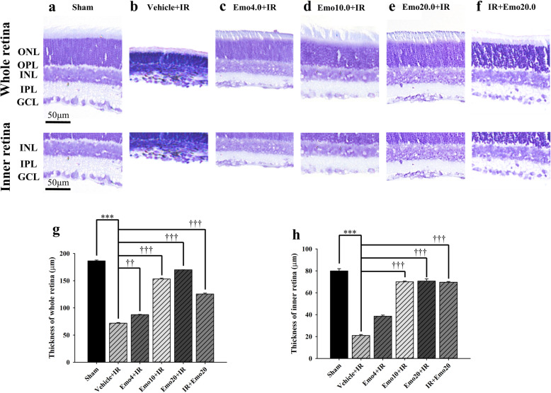
Fig. 3.
Calculation of thickness of cresyl violet stained retinas. a ~ f. Cresyl violet stained retinal sections with the same eccentricity. Micrographs showing the whole (top row) or inner retina (bottom row) thickness (μm) of different groups. a, b, g and h. As compared with the retinal thickness of the control group (Sham: 186.50 ± 1.43; inner = 79.90 ± 2.06), a substantial reduction in the whole or inner retina thickness was observed in that of the Vehicle+IR group (whole = 71.80 ± 1.08; inner = 20.97 ± 0.85). c ~ e, g and h. Pre-ischemic intravitreous injection of emodin dose-responsively (least effect at 4 μM; Emo4 + IR: whole = 87.40 ± 0.60, inner = 38.60 ± 1.01; then, 10 μM; greatest effect at 20 μM) and in a significant way (Emo10 + IR: whole = 153.20 ± 1.48, inner = 70.05 ± 0.60; Emo20 + IR: whole: 170.10 ± 0.10; 70.65 ± 2.06) attenuated ischemia induced reduction in the whole and inner retina thickness. f, g and h. Post-ischemic intravitreous injection of 20 μM emodin also significantly alleviated ischemia reduced whole and inner retina thickness (IR + Emo20: whole = 125.45 ± 1.68, inner = 69.65 ± 0.68). g and h. Quantitative analysis of the whole or inner retina thickness. *** or †††/†† indicates a significant (P < 0.001 or P < 0.001/P < 0.01) difference from the Sham or Vehicle+IR group. Abbreviations: IR, ischemia plus reperfusion; ONL, outer nuclear layer; OPL, outer plexiform layer; INL, inner nuclear layer; IPL, inner plexiform layer; GCL, ganglion cell layer. Scale bar = 50 μm. The results are mean ± standard error. Abbreviations for group names are provided in Table 3

