
Figure 5. AIM2 inflammasome contributes to pancreatic tumorigenesis.
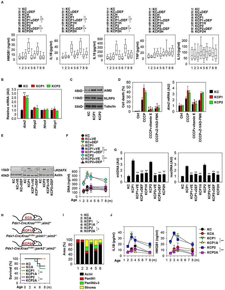
(A) Analysis of cytokines in serum in the indicated mice with or without deferiprone (“DEF”) treatment at three months of age (n=10 mice/group, * p < 0.05, unpaired t-test, data are expressed as median value [black line], interquartile range [box], and minimum and maximum of all data [black line]). (B) Analysis of expression of the indicated genes in pancreata from in KC, KCP1, and KCP2 mice at three months of age (n=3 mice/group, ** p< 0.01 versus KC group, unpaired t-test, data are expressed as means ± s.e.m., AU, arbitrary units.). (C) Analysis of the expression of indicated proteins in pancreata from in KC, KCP1, and KCP2 mice at three months of age. (D) PDAC cells from KC, KCP1, or KCP2 mice were treated with CCCP (10 μM) in the absence or presence of vitamin E (50 μM) or Z-VAD-FMK (20 μM) for 24 h, and the levels of cell death and Aim2 mRNA were assayed by CCK-8 kit and Q-PCR, respectively (n=3, * p < 0.05 versus CCCP group, unpaired t-test, data are expressed as means ± s.e.m., AU, arbitrary units.) (E) Analysis of pancreatic γH2AFX expression from KC, KCP1, and KCP2 mice with or without vitamin E (“VE”) or deferiprone (“DEF”) treatment at indicated ages. (F) Analysis of serum DNA levels from KC, KCP1, and KCP2 mice with or without vitamin E (“VE”) or deferiprone (“DEF”) treatment at indicated ages (n=3 mice/group, ** p < 0.01, unpaired t-test, data are expressed as means ± s.e.m.). (G) Analysis of indicated DNA levels in KC, KCP1, KCP2, KCP1H, and KCP2H mice with or without vitamin E (“VE”) or deferiprone (“DEF”) treatment at three months of age (n=5 mice/group, ** p < 0.01, unpaired t-test versus KCP1 or KCP2 group, data are expressed as means ± s.e.m.). (H) Kaplan-Meier survival analysis was performed with the indicated mice (n=10-20 mice/group, ** p < 0.01, log-rank test). (I) Histological evaluation of pancreata at three months of age (n=5 mice/group, * p < 0.05, unpaired t-test, data are expressed as percentage of positive area). (J) Analysis of the indicated serum cytokine levels in mice (n=3 mice/group, ** p < 0.01, ANOVA test, data are expressed as means ± s.e.m.). See also Figures S4 and S5.