Figure 3.

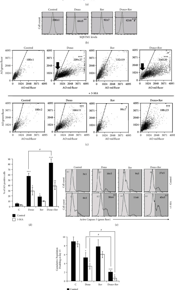
Rsv potentiated cytoprotective autophagy induced by Doxo in MCF7 cells. (a) Experimental design. Cells were treated with Rsv 30 μM, Doxo 100 nM, or Rsv30+Doxo100 for 24 h. Dimethyl Sulfoxide (DMSO) not exceeding 0.05% was used as control. After this, cell viability was assessed. Then, cells were replated in a Drug-Free Medium and grown for 15 days. Cells were treated with 2 mM of 3-methyladenine (3-MA) for 1 h at days 3 and 4. (b) SQSTM1 levels measured by immunocytochemistry at day 5 (average ± standard deviation). (c) Acridine orange (AO) staining. Numbers represent the intensity of AO red fluorescence in relation to control, considered as 100 (average ± standard deviation). Black arrow points to the AO-negative population of cells in Doxo and Doxo+Rsv treatment. (d) Percentage of AO-positive cells. (e) Active caspase-3-positive cells measured by flow cytometry. Numbers correspond to the percentage of positive cells (average ± standard deviation). (f) Cumulative Population Doubling measured at day 15. Abbreviations: AO: acridine orange; SQSTM1: sequestosome 1; 3-MA: 3-methyladenine; Casp-3: active caspase-3-positive cells; CPD: Cumulative Population Doubling. ∗p < 0.05, ∗∗p < 0.01, and ∗∗∗p < 0.001 in relation to control; #p < 0.05, ##p < 0.01, and ###p < 0.001 in relation to Doxo; †p < 0.05, ††p < 0.01, and †††p < 0.001, comparing 3-MA to control using PBS.
