Figure 3.

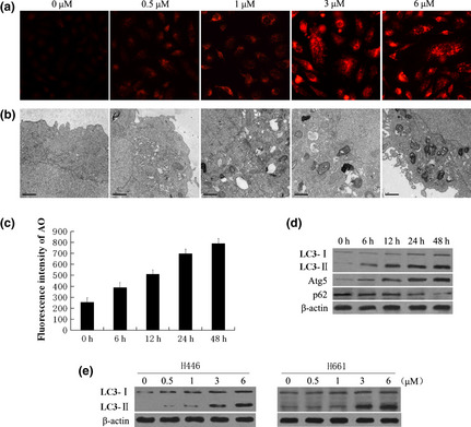
Induction of autophagy in a variety of human lung cancer cells in response to peneciraistin C (Pe‐C). (a, b) A549 cells were treated with indicated concentrations of (Pe‐C) for 48 h. (a) Treated cells were stained with acridine orange (AO) and visualized under a red filter fluorescence microscope. (b) Formation of autophagosomes in treated cells was checked by transmission electron microscopy. Scale bar = 1 μM. (c, d) A549 cells were treated with 3 μM Pe‐C for the indicated times. (c) Treated cells were stained with AO, then the fluorescence intensity was analyzed by flow cytometry. (d) After treatment, whole cell extracts were subjected to immunoblotting with anti‐LC3, anti‐Atg5, and anti‐p62 antibodies. (e) H446 and H661 cells were treated with indicated concentrations of Pe‐C for 48 h. The LC3‐I/LC3‐II levels were determined as described in (d).
