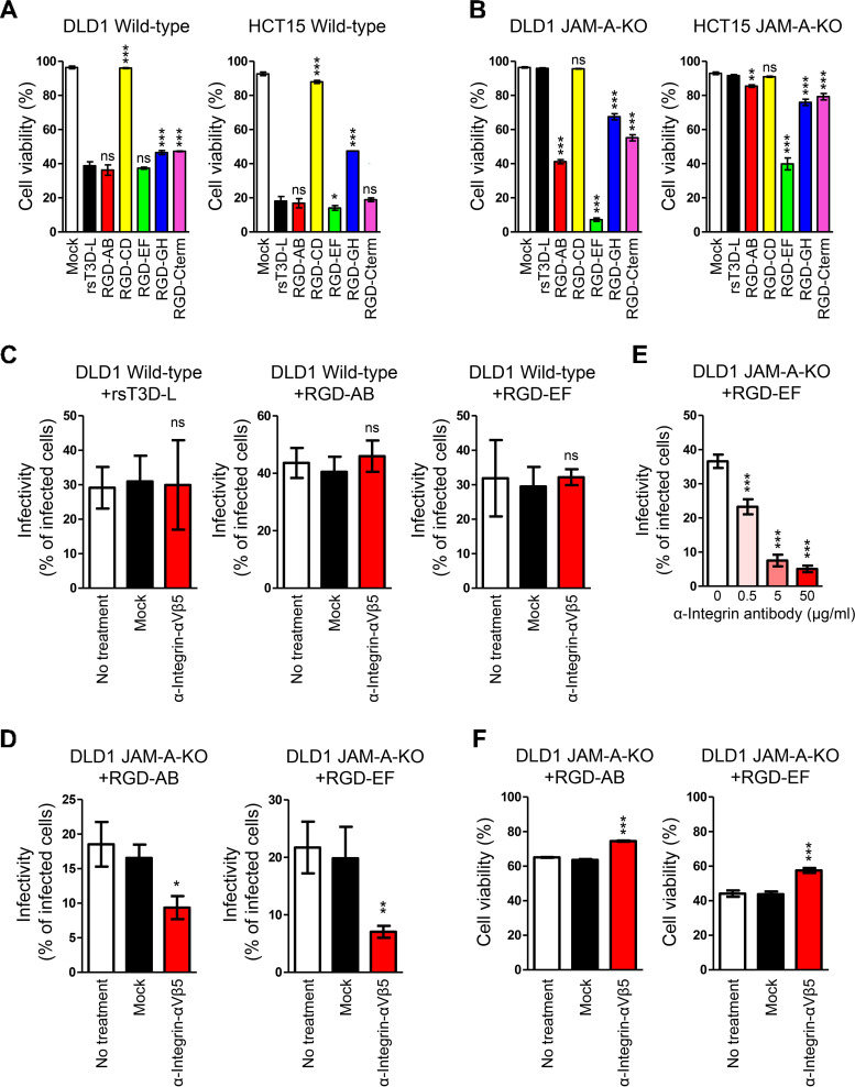
FIG 5.
Integrin-dependent oncolysis of JAM-A knockout cell lines by RGD σ1-modified viruses. (A and B) Oncolytic activity of RGD σ1-modified viruses in cell lines. Wild-type (A) and JAM-A-KO (B) cells were infected with wild-type and RGD σ1-modified viruses at an MOI of 10 PFU/cell. Cells were collected every 24 h and cell viability was measured by propidium iodide staining. Each value represents an average from triplicate samples. The error bars indicate the standard deviation. Representative results from three independent experiments are shown. Significant differences were determined using one-way ANOVA; *, P < 0.05; ***, P < 0.0001; ns, not significant. (C and D) Infection of cancer cells by RGD σ1-modified viruses following treatment with anti-integrin antibodies. Wild-type DLD1 (C) and DLD1 JAM-A-KO (D) cells were incubated at 37°C for 1 h with an anti-integrin αVβ5 antibody (P1F6) or an anti-FLAG antibody and then infected for 16 h with rsT3D-L, RGD-AB, or RGD-EF at an MOI of 50 PFU/cell. Infectivity was analyzed by indirect immunofluorescence analysis using an anti-T3D antibody. Infectivity was calculated as the ratio of infected cells to the total number of cells. The results are expressed as the mean score for four independent fields of view. The error bars indicate the standard deviation. Representative results from two independent experiments are shown. Significant differences were determined using Student’s t test; *, P < 0.05; **, P < 0.001; ns, not significant. (E) Infection of JAM-A-KO cells by RGD-EF following treatment with different concentrations of an anti-integrin antibody. DLD1 JAM-A-KO cells were incubated at 37°C for 1 h with an anti-integrin αVβ5 antibody (P1F6) or an anti-FLAG antibody, and then infected for 16 h with RGD-EF at an MOI of 50 PFU/cell. Infectivity was analyzed by indirect immunofluorescence analysis using an anti-T3D antibody. Infectivity was calculated as the ratio of infected cells to the total number of cells. The results are expressed as the mean score for four independent fields of view. The error bars indicate the standard deviation. Representative results from two independent experiments are shown. Significant differences were determined using one-way ANOVA; ***, P < 0.0001. (F) Oncolysis of RGD σ1-modified viruses following treatment with an anti-integrin antibody. DLD1 JAM-A-KO cells were incubated at 37°C for 1 h with an anti-integrin αVβ5 antibody (P1F6) or an anti-FLAG antibody, and then infected for 16 h with RGD-AB or RGD-EF at an MOI of 50 PFU/cell. Cell viability was measured by propidium iodide staining. Each value represents an average from triplicate samples. The error bars indicate the standard deviation. Representative results from two independent experiments are shown. Significant differences were determined using Student’s t test; ***, P < 0.0001.

