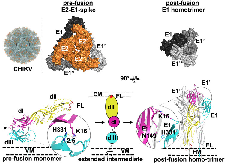
FIG 1.
CHIKV envelope proteins pre- to postfusion conformation switch. (Top, left to right) Image from 3D reconstruction of CHIKV E1-E2 into CHIKV virus-like particles (EMDB ID 5577); the surface view (labeled “CHIKV”) is shown. One trimeric spike structure is shown in the circle. An enlarged image of one E2-E1 trimeric spike is shown to the left (PDB ID 3J2W). Top views of E1 (different shades of gray) and E2 (orange) are shown in a surface representation. Three protomers of E1 and E2 are labeled E1, E′, and E′′ and E2, E2′, and E2′′, respectively. The top view of the postfusion E1 homotrimer (PDB ID 1RER) is shown to the right. (Bottom left) A single monomer of E1 (from PDB ID 3N42), in the prefusion conformation, is shown in cartoon representation with the three domains (dI, dII, and dIII) and fusion loops (FL) labeled. The dI-III linker is marked with a black arrow. The middle and right portions show a schematic of the extended intermediate (middle [dIII, dI, and dII and fusion loops depicted in cyan, magenta, and yellow ovals and a red star, respectively]) and postfusion (right) E1 homotrimer conformation. The domain I-III interface His 331-Lys 16 H bond in prefusion and the His 331-Asn 149 H bond in postfusion are shown in a cartoon and stick representation with enlarged areas in circles. VM, CM, and FM refer to viral membrane, cell membrane, and fused membrane, respectively. Dashed lines connecting protein structure figures with VM represent the stem and transmembrane region that anchor the proteins into VM or FM.

