Fig. 2.
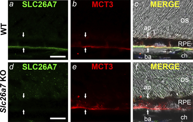
Immunolocalization of SLC26A7 in the mouse retinal pigment epithelium (RPE) basolateral membrane. Confocal and DIC images of retinal cryosections from wild-type (WT) mice (a–c) and Slc26a7 knockout (KO) mice (d–f). SLC26A7 immunostaining colocalized with the RPE basolateral membrane marker MCT3 in WT mouse RPE (c) but was absent in Slc26a7 KO mouse RPE (d). Scale bars in a and d, 10 µm; ap, apical membrane; ba, basolateral membrane; ch, choroid; os, photoreceptor outer segments. Downward and upward arrows point to the apical and basal aspects of the RPE, respectively. Retinal cryosections were immunostained with a mouse monoclonal anti-SLC26A7 antibody (17) and rabbit polyclonal anti‐MCT3 antibodies (30) followed by Alexa Fluor 448- and 555‐tagged secondary antibodies. Laser power and photomultiplier tube gain for the 488-nm (green) channel were identical in a and d. The immunolocalization of SLC26A7 to the basolateral membrane shown in a is representative of results from 5 independent experiments on retinal sections from three 3–6-wk-old WT mice of both sexes.

