Fig. 3.
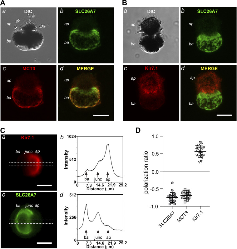
Isolated mouse retinal pigment epithelial (RPE) cells retain the polarized expression of SLC26A7 in the basolateral membrane. A and B: confocal and DIC images of fixed, isolated wild-type (WT) mouse RPE cells double labeled with anti-SLC26A7 and anti-MCT3 (A) or anti-Kir7.1 (B) antibodies. Scale bars, 10 µm; ap, apical pole; ba, basolateral pole. The results for SLC26A7, MCT3, and Kir7.1 immunolabeling are representative of 40 cells from 4 adult WT mice, 42 cells from 2 adult WT mice, and 33 cells from 2 adult WT mice, respectively. C: wide-field epifluorescence images of a fixed WT mouse RPE cell double labeled with anti-Kir7.1 (a) and anti-SLC26A7 (c) antibodies. Note that the orientation of the cell differs from that in B. The dashed horizontal lines mark the region subjected to line scan analysis, the results of which are shown in b (Kir7.1) and d (SLC26A7). Scale bars, 10 µm; junc, junction. D: quantification of the membrane polarity of SLC26A7, MCT3, and Kir7.1 expression in isolated mouse RPE cells from line scan analysis of fluorescence along the major axis as in C, b and d, using Eq. 1. Positive values indicate predominantly apical membrane expression, whereas negative values indicate predominantly basolateral membrane expression. Symbols represent measurements in individual cells (n = 21 cells for each group), and horizontal lines and error bars represent means ± SD. The cells used in this analysis were isolated from two adult WT mice.

