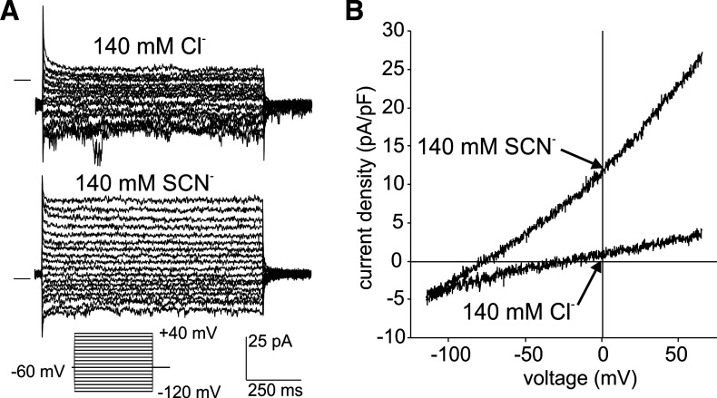
Fig. 8.
Macroscopic anion currents recorded from an excised outside-out basolateral membrane patch from an Slc26a7 knockout (KO) mouse retinal pigment epithelium cell. A: representative current recordings from a basolateral membrane patch evoked by voltage steps in the range −120 to +40 mV from a holding potential of −60 mV in the presence of 140 mM external NaCl (top) or 140 mM external sodium thiocyanate (NaSCN; bottom). The horizontal lines to the left of the traces indicate the zero-current level. The pipette contained 140 mM N-methyl-d-glucamine (NMDG)-Cl solution. Replacement of Cl− with thiocyanate (SCN−) increased the amplitude of outward currents. B: current-voltage plots of currents evoked by voltage ramps in the same patch in the presence of 140 mM external NaCl or 140 mM external NaSCN. The membrane potential was held at −60 mV and ramped from −120 mV to +60 mV over a period of 1 s. Replacement of Cl− with SCN− increased the outward conductance from 40.8 pS/pF to 269.7 pS/pF and shifted the reversal potential from −20.0 to −75.8 mV.

