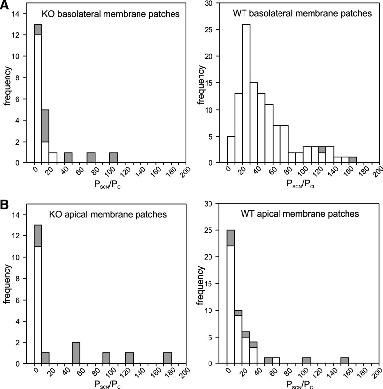
Fig. 10.
Deletion of Slc26a7 decreases the relative thiocyanate (SCN−) permeability (PSCN/PCl) of the mouse retinal pigment epithelium (RPE) basolateral membrane. A: histograms showing the distribution PSCN/PCl values for outside-out basolateral membrane patches from Slc26a7 knockout (KO) mouse (left) and wild-type (WT) mouse (right) RPE cells. Not shown in the histogram for WT mouse RPE basolateral membrane patches are PSCN/PCl values of 308.1, 309.2, 357.1, 366.3, and 519. The mean values of PSCN/PCl in Slc26a7 KO mouse RPE basolateral membrane patches [17.4 ± 5.4 (SE), n = 22 cells from 14 mice of both sexes, 3.9–40.0 wk old] and WT mouse RPE basolateral membrane patches [63.8 ± 6.8 (SE), n = 121 cells from 47 mice of both sexes, 4.9–19.9 wk old] were significantly different (P < 0.001, 2-tailed Mann–Whitney test). Open regions of bars indicate data obtained at a holding potential of −60 mV, and gray regions indicate data obtained at a holding potential of −120 mV. B: histograms showing the distribution of PSCN/PCl values for outside-out apical membrane patches from Slc26a7 KO mouse (left) and WT mouse (right) RPE cells. Not shown in the histogram for WT mouse RPE apical membrane patches are PSCN/PCl values of 263.7, 280.7, and 703.0. The mean values of PSCN/PCl Slc26a7 KO mouse RPE apical membrane patches [30.8 ± 11.7 (SE), n = 19 cells from 11 mice of both sexes, 3.6–10.6 wk old] and WT mouse RPE apical membrane patches [42.6 ± 15.1 (SE), n = 52 cells from 25 mice of both sexes, 3.6–14.4 wk old] were not significantly different (P = 0.10, 2-tailed Mann–Whitney test). Data for WT RPE cells are from our previously published study on Slc26a7+/+ mice (10).

