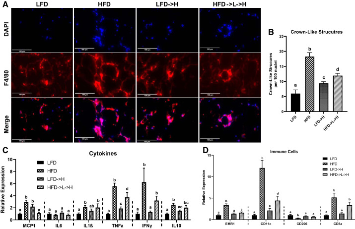
Fig. 3.
Epididymal fat gene expression indicates inflammatory memory may exist in weight-cycling mice. A: representative ×40immunofluorescence staining images of macrophage marker F4/80 in epididymal adipose tissue shows significant crowning in HFD and HFD→L→H mice. B: quantification of crown-like structures per 100 nuclei. C: gene expression of proinflammatory cytokines MCP1, IL-6, IL-1β, TNFα, IFNγ, and anti-inflammatory cytokine IL-10. D: gene expression of total macrophage marker EMR1, M1 macrophage marker CD11c, M2 macrophage marker CD206, and cytotoxic T cell marker CD8a. LFD, low-fat diet for 32 wk; HFD, high-fat diet for 32 wk; LFD→H, LFD for 28 wk and then changed to a HFD for 4 wk; HFD→L→H, HFD for 21 wk, then changed to LFD for 7 wk, and then changed to HFD for 4 wk. Data are shown as means ± SE (n = 8–10/group). Bar graphs not sharing a common letter are significantly different from one another according to one-way ANOVA (P < 0.05).

