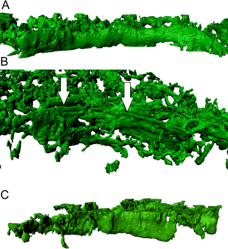
Fig 2. Morphological appearance of alpha-actin positive fibroblastic reticular cell (FRC) walls in lymphadenitis (LAD).
(A) 3D view from the germinal centre towards the T-zone shows clear B/T-border compartmentalisation by FRC walls in lymphadenitis. This image contains a consistent FRC wall defining a clear barrier between two lymph node compartments. Arrows (in white) point on an FRC wall. (B) The point of view of this image is from the germinal centre towards the bordering T-zone, depicting a FRC wall. (C) FRC wall with a clearly defined border towards B-zone. The surface is smoother and more regular in comparison to the alveolate structure within the T-zone. The maximum resolution of (A), (B) and (C) was set to 0.13 μm per pixel in the native microscopy dataset.

