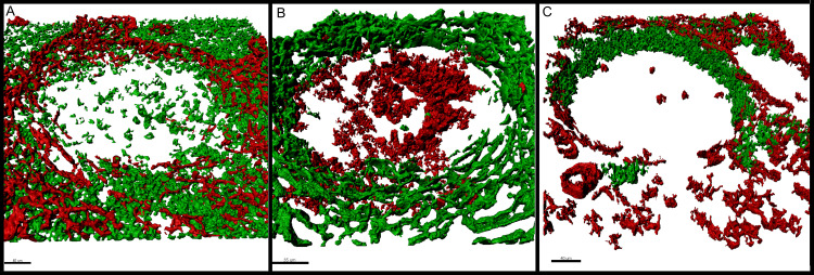
Fig 3. Description of the T-zone, the follicular mantle and the germinal centre in relation to the physical FRC boundary.
(A) Alpha-actin+ (red) and CD3+ cells (green) relate to each other. The number of T-cells decreases abruptly towards the B-zone where the FRC wall is located. (B) Alpha-actin positive (green) and BCL6 positive (red) cells show proportion of walls towards the germinal centre. BCL6 positive germinal centre cells are situated within the physical boundary. Additionally, the BCL6 and alpha-actin negative surrounding between germinal centre and FRC wall indicates that the follicular mantle is within the compartmentalised area of the walls. (C) Alpha-actin-positive (red) and IgD positive (green) structures reveal orientation of FRC walls. The follicular mantle shows a polar orientation without or with only fragmented FRC walls towards the mantle zone. On the opposite pole, the germinal centre is compartmentalised by smaller FRC walls and is more permeable. The remaining germinal centre is enclosed from both sides by the FRC walls in form of a sufficient physical boundary. This staining combination shows properly that the orientation of the FRC walls shows a coherence with the follicular mantle. The maximum resolution of (A), (B) and (C) was set to 0.13 μm per pixel in the native microscopy dataset.

