
Fig 4. Morphological appearance of alpha-actin positive fibroblastic reticular cell (FRC) walls in angioimmunoblastic T-cell lymphoma (AITL).
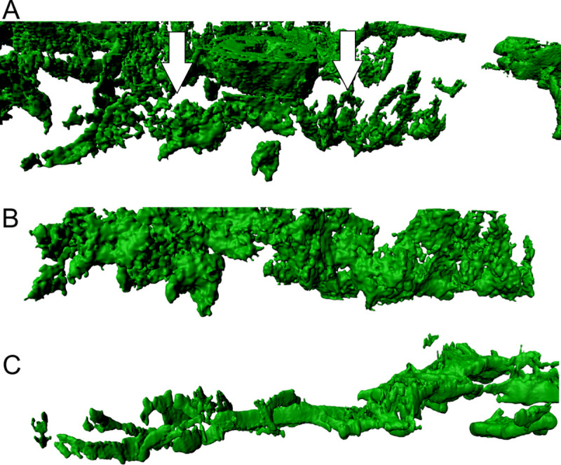
(A) Walls in AITL present themselves as less dense and not consistent as shown by the arrows (in white). The surface is inconsistent and permeable. (B and C) The FRC walls shown here display a rougher surface in comparison to LAD. In addition to that, they are more scattered and of variable morphology. The maximum resolution of (A), (B) and (C) was set to 0.13 μm per pixel in the native microscopy dataset.