
Figure 1.
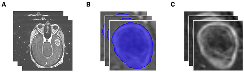
Preprocessing Workflow. (A) Input: slices of pre-treatment T1-post contrast brain MRI scans. (B) Identification of the region of interest from manual segmentations. (C) Output: extracted tumors with pixel resampling, N4ITK bias field correction, and z-score normalization.