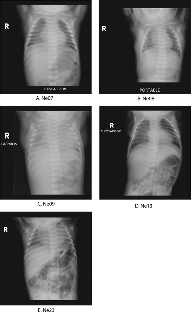
FIGURE 3.

Chest radiograph images of 5 neonates with SARS-CoV-2 infections. A: Chest radiograph of Ne07 showed patchy opacities in right lower lobe suggesting pneumonia. B: Chest radiograph of Ne08 looked normal. C: Chest radiograph of Ne09 showed bilateral ground-glass opacity indicative of SARS-CoV-2. D: Chest radiograph of Ne13 showed few patchy opacities in the right lower perihilar region indicating nonspecific inflammatory lesion in right lower zone. E: Chest radiograph of Ne23 looked normal.
