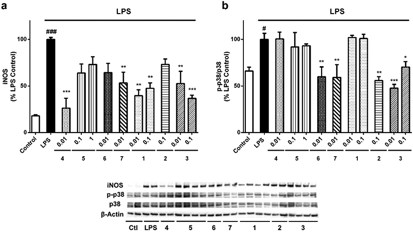
Figure 5.

Effects of gracilin A derivatives on iNOS and p38 MAPK. Microglial cells were treated with compounds at μM concentrations and 500 ng/mL LPS. Then, BV2 cells were lysed and the protein expression levels of (a) iNOS and (b) p38 were analysed by western blot. The activation of the kinase was determined as the ratio between phosphorylated and total protein levels. Protein expression levels were corrected by β-actin. Treatments are reordered to match with blots. Values are mean± SEM of three independent experiments carried out by duplicate. Treatments with derivatives are compared to LPS control cells by one way ANOVA and Dunnett’s test (*p<0.05, **p<0.01, ***p<0.001). Cells treated with LPS alone are compared to untreated control cells (#p<0.05, ###p<0.001)
