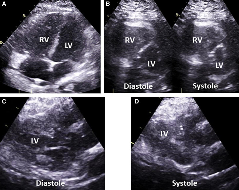
Figure 1.

Examples of right ventricular (RV) and left ventricular (LV) echocardiographic findings. Transthoracic echocardiographic images are shown for each. A, RV dilation is demonstrated by a diastolic area equal to, or larger than, the LV diastolic area. B, Abnormal septal motion including flattening of the interventricular septum and displacement of the septum throughout the cardiac cycle demonstrating elevated RV pressures. C, Underfilled LV with a LV internal end-diastolic diameter of less than 4.0 cm. D, Hyperdynamic LV with ejection fraction greater than 60% seen here with near complete obliteration of the LV cavity.
