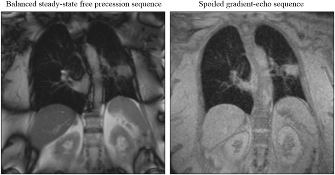
Figure 6.
Two coronal 2D cine MR images of a lung cancer patient acquired with different gradient-echo MR sequences. One has been acquired with a balanced steady-state free precession sequence providing a T2/T1-weighted contrast, while the other was obtained using a spoiled gradient-echo sequence with a T1-weighted contrast (Menten et al, unpublished).

