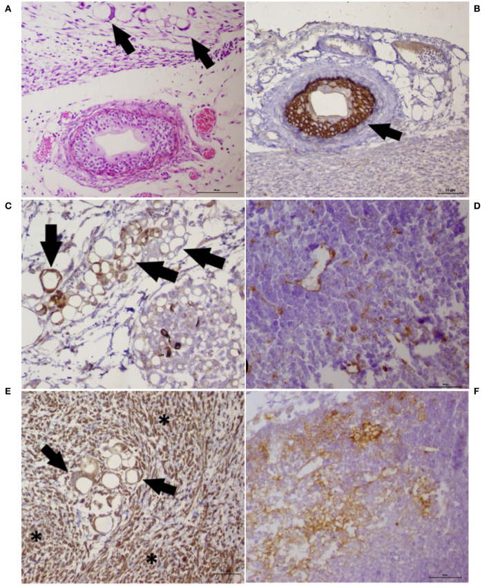
Figure 6.
Histochemistry and immunohistochemistry analyses of a tumor growth (xenograft) from the cancer cell lines UNESP-MM4 and UNESP-CM60. (A) HE staining showing neoplastic cells forming vascular-like structures (arrows) and the presence of a blood vessel showing tumor cells growing into the blood vessel from the UNESP-MM4 cell line. (B) Positive pancytokeratin expression in the neoplastic cells growing inside a blood vessel (arrow) from the UNESP-MM4 cell line. (C) Positive cytokeratin 8/18 expression of neoplastic cells forming vascular-like structures and confirming its epithelial origin. (D) Vascular-like structure from the xenotransplantation of the UNESP-CM60 cell line. Note the scattered cytokeratin 8/18 expression of neoplastic cells, including vascular-like structures, which confirms its epithelial origin. (E) Vimentin expression of tumor growth from the UNESP-MM4 cell line. Note the positive vimentin expression in neoplastic (arrows) and stromal (asterisk) cells. (F) Vimentin-positive expression in tumor growth from xenotransplantation of the UNESP-CM60 cell line.

