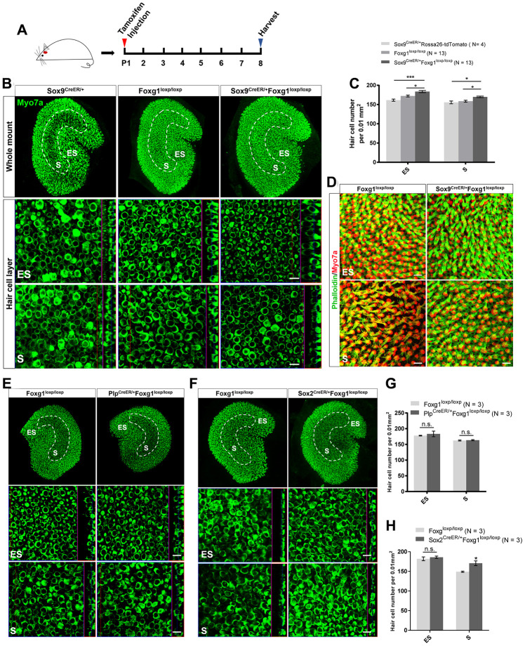
Figure 2.
cKD of Foxg1 in Sox9+ SCs led to increased HCs number in the mouse utricle at P08. (A) Tamoxifen was injected into P01 mice to knock down Foxg1 in Sox9+ SCs, and the utricle was harvested at P08. (B) Immunofluorescence staining with anti-Myo7a (green) antibodies in the utricle of P08 Sox9CreER/+, Foxg1loxp/loxp, and Sox9CreER/+Foxg1loxp/loxp cKD mice. (C) Quantification of HCs in the ES and S regions of the utricle. *p < 0.05, ***p < 0.001. (D) Immunofluorescence staining with phalloidin (green) in the utricle of P08 Foxg1 cKD and control mice. (E, F) Immunofluorescence staining with anti-Myo7a (green) antibodies in the utricle from P8 PlpCreER/+Foxg1loxp/loxp (E) and Sox2CreER/+Foxg1loxp/loxp mice (F). Foxg1loxp/loxp mice were used as the control mice. (G, H) Quantification of the total numbers of HCs in the ES and S regions of the utricle from the above mice. *p < 0.05, n.s. not significant. For all experiments, Myo7a was used to indicate the HC. Scale bar, 10 μm. N indicates the number of mice, data are represented as mean ± SEM.

