Figure 3.
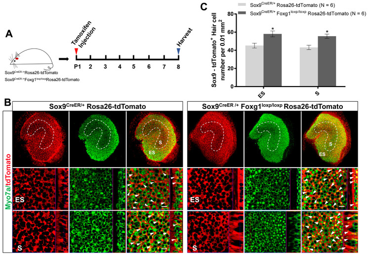
Foxg1 cKD led to increased trans-differentiation of Sox9+ SCs in the utricle in vivo. (A) P01 mice were i.p. injected Tamoxifen for activating the Cre enzyme, and the mice were sacrificed at P08. (B) Immunofluorescence staining with anti-Myo7a (green) antibodies in the utricle from P08 Sox9CreER/+Rosa26-tdTomato and Sox9CreER/+Foxg1loxp/loxpRosa26-tdTomato mice. Myo7a was used as the HC marker. tdTomato+ HCs are indicated by white arrows. (C) Quantification of tdTomato+ HCs per 0.01 mm2 area in both the S and ES regions of the P08 mouse utricle. Scale bar, 10 μm. N indicates the number of mice. *p < 0.05, data are represented as mean ± SEM.

