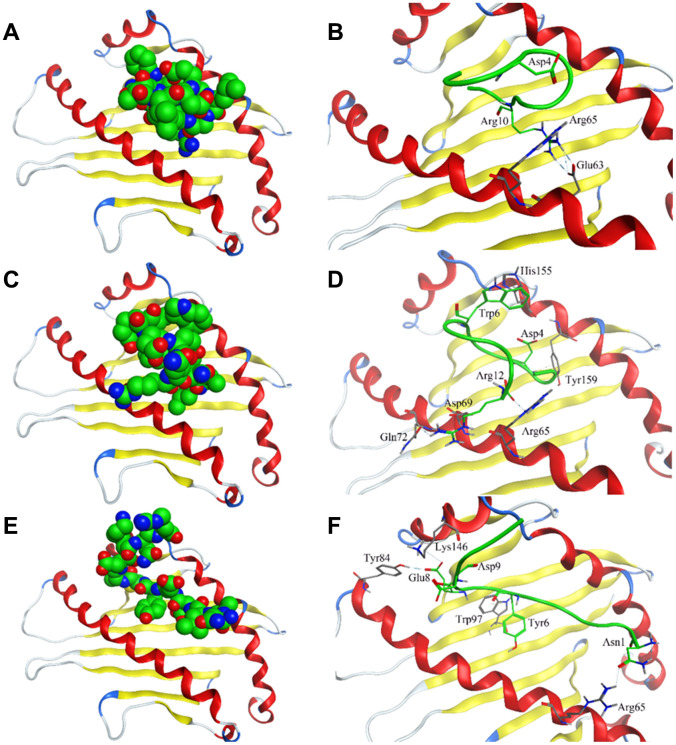
Figure 4.
Interaction of affinity peptides with HLA-E. (A, C, E). The docking of PEPTIDE 1, 2 and 3 with the key area of HLA-E (PDB ID: 3CDG). (B, D, F). Site view of PEPTIDE 1, 2 and 3 docking with HLA-E. Dash lines, hydrogen bonds; Labeled residues, amino acids interacted between affinity peptides and HLA-E.

