
Fig 2.
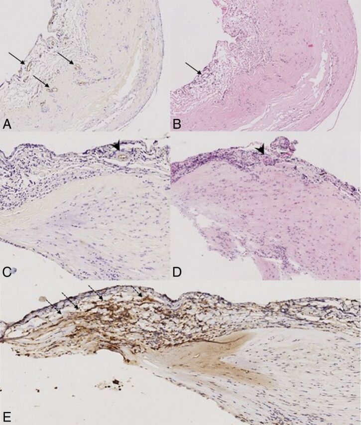
CD34 (A and C), hematoxylin-eosin (B and D), and MPO stain (E) at 10x magnification. A and B, Patient 3: aneurysm wall with evidence of neovascularization (arrows). C and D, Patient 8, vasa vasorum are present (arrowheads). There is no sign of neovascularization. E, The same patient as in C and D. MPO-positive aneurysm wall with accumulation of MPO-positive inflammatory cells in the tunica media (dotted arrows).