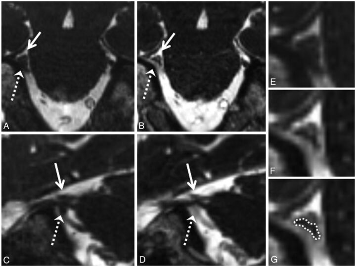
Fig 2.
Grade 2 neurovascular conflict. Coronal NE-CISS (A) and CE-CISS (B) and sagittal NE-CISS (C) and CE-CISS (D) images show neurovascular conflict of the cisternal segment of the patient's right trigeminal nerve with a branch of the superior cerebellar artery from above (solid white arrow) and the superior petrosal vein from below (dashed white arrow), resulting in flattening of the nerve near the porus trigeminus. On the NE-CISS images (A and C), the nerve is not well-delineated from the adjacent vascular structures. On the CE-CISS images (B and D), the vessels enhance, outlining the compressed nerve between them. Zoomed-in images of the site of neurovascular conflict in the coronal plane (E and F) illustrate the poor contrast between vessels and nerve on the NE-CISS image (E) and the improved contrast after administration of gadolinium contrast material (F), allowing more confident delineation of the compressed nerve from the adjacent vessels (G). Both NE-CISS and CE-CISS images were interpreted as grade 2 compression. On the patient's left, grade 0 was given for both the NE-CISS and CE-CISS images.

