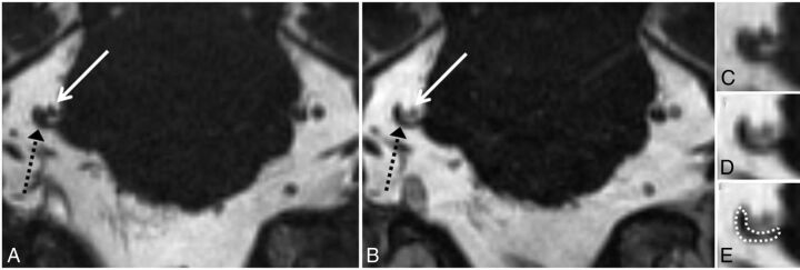
Fig 3.
Grade 3 neurovascular conflict. Coronal NE-CISS (A) and CE-CISS (B) images show compression of the cisternal segment of the patient's right trigeminal nerve (black dashed arrow) by branches of the superior cerebellar artery from above (solid white arrow). On the unenhanced image (A), the nerve root is not well-distinguished from the compressing arterial branches. After contrast administration (B), at least 2 arterial branches enhance, allowing more confident delineation of the markedly compressed nerve. Zoomed-in images of the site of neurovascular conflict in the coronal plane (C–E) illustrate the poor contrast between vessels and nerve on the NE-CISS image (C) and the improved contrast after administration of gadolinium contrast material (D), allowing more confident delineation of the compressed nerve from the adjacent vessels (E). In this patient, grade 3 was given for both the NE-CISS and CE-CISS images, but the measured CSA was lower and the degree of flattening was more pronounced on the CE-CISS images. On the patient's left, grade 0 was given for both the NE-CISS and CE-CISS images.

