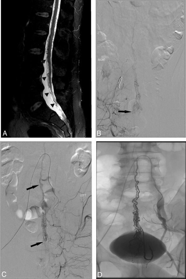
Fig 1.
A, Sagittal T2-weighted spine MR imaging showing tethering of the spinal cord (lower arrowheads). Note the conus tip at the L4–L5 level and flow voids throughout the spinal canal, most prominent in the sacral canal (arrowheads), extending superiorly to approximately the T2 level. B, Right internal iliac artery angiogram demonstrates an arteriovenous shunt in the spinal canal of the sacrum (black arrow). The feeding artery enters through the left S4 neural foramen. C, Left internal iliac artery angiogram demonstrates an additional feeder to the arteriovenous shunt (lower black arrow). Intradural draining veins (upper black arrow) are visible. D, Angiogram obtained following embolization demonstrates a large cast of Onyx extending from the site of fistula to the coronal venous plexus.

